Dental x-rays (also called radiographs) are a valuable part of dental treatment because they can detect damage to teeth and gums not visible during a routine visual examination.
For example, x-rays can show the condition of your teeth, their roots, jaw placement and the overall composition of your facial bones. X-rays can help your dentist determine the presence or degree of periodontal (gum) disease, cavities, abscesses and many abnormal growths, such as cysts and tumors. X-rays also can show the exact location of impacted teeth and teeth that have not yet fully developed. Finding and treating dental problems at an early stage can save time, money and unnecessary discomfort. If you have a hidden tumor, x-rays may even help save your life.
X-RAY FAQ
 Do all patients have x-rays taken every six months?
No. Your x-ray schedule is based on the dentist’s assessment of your individual needs, including whether you’re a new patient or a follow-up patient, adult or child.* Dentists typically require new patients to have a full set of mouth x-rays in order to evaluate oral health status, including any underlying signs of gum disease, and for future comparison. (If you change dentists and have recently had x-rays taken, ask to have them sent to the new dentist.) Dentists may require x-rays during follow-up visits to monitor oral health.
What kind of x-rays does my dentist usually take?
The most common type of x-rays dentists take are known as bitewing x-rays. These require patients to hold or bite down on a piece of plastic with x-ray film in the center. Bitewing x-rays typically determine the presence of decay in-between teeth — one of the most common areas where decay-causing bacteria reside.
Another common dental x-ray type is the periapical x-ray, which gives the dentist an image of the entire tooth, including the roots. With periapical x-rays, dentists evaluate a particular tooth’s root structure and bone level and also can detect cysts and abscesses.
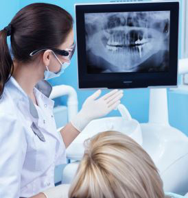
My dentist has ordered a “panoramic radiograph.” What is that?
Just as a panoramic photograph allows you to see a broad view of a large area, such as the Grand Canyon, a panoramic x-ray (also called radiograph) allows your dentist to see the entire structure of your mouth (all of your upper and lower teeth and parts of your jaw) in a single image. A common use of a panoramic x-ray, for example, is to assess teeth development in a child or teenager, especially wisdom teeth. Also known as third molars, wisdom teeth don’t erupt until the teenage years or beyond and can cause crowding or become impacted (because the teeth don’t have enough room to grow). A panoramic x-rays is also used to help evaluate dental implant placements.
Why would I need more than one type of x-ray?
What is apparent through one type of x-ray often is not visible on another. The panoramic x-ray will give your dentist a general and comprehensive view of your entire mouth on a single film, which a bitewing or periapical x-ray cannot show. On the other hand, periapical or bitewing x-rays show a detailed image of a smaller area, making it easier for your dentist to see decay or cavities between your teeth. X-rays are not prescribed indiscriminately. Your dentist has a need for the different information that each radiograph can provide to formulate a diagnosis.
Should I be concerned about exposure to radiation?
All health care providers are sensitive to patients’ concerns about exposure to radiation. Your dentist has been trained to prescribe x-rays when they are appropriate and to tailor their frequency to each patient’s individual needs. By using state-of-the-art technology and staying knowledgeable about recent advances, your dentist knows which techniques, procedures and x-ray films can minimize your exposure to radiation. Dr. Koshki has used only digital x-ray technology since 2009 which ensures the very least amount of exposure.